| Endotracheal
tubes
Tracheal
intubation is simple in most domestic species of animal, so
an endotracheal tube is almost invariably used in preference
to a mask for connection of the breathing circuit to the patient's
airway.
Benefits
of endotracheal intubation
- Assists
in maintaining a patent airway.
- Prevents
aspiration of saliva or regurgitated gastric contents.
- Seals the
respiratory system and breathing circuit.
Types
of endotracheal tube
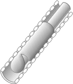
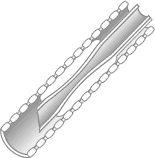
Magill tube
Magill tubes are those most commonly used in veterinary anesthesia.
They are curved and have a bevelled tip which facilitates passing
the tube through the larynx. They are available in a variety of
sizes from 3 mm to 40 mm internal diameter (the internal diameter,
ID, being the standard method of referring to the size of tube),
accommodating animals ranging in size from cats to large horses.
 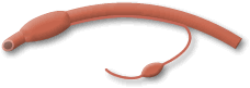
Magill tubes
are manufactured either plain or with an inflatable cuff. Plain
tubes are used in veterinary anesthesia when the volume of the cuff
would hinder insertion of a tube of sufficient internal diameter,
as in very small animals (e.g. cats and birds).
Two types of
cuff are commonly used. The traditional high pressure, low volume
cuff operates by distending a rubber balloon around the tip of the
tube, thus sealing the trachea. This carries the risk that the pressure
within the cuff can cause trauma to or necrosis of the tracheal
wall.
The more modern
low pressure, high volume cuff has a much greater volume than the
traditional cuff, and so requires a lower inflation pressure to
produce a seal. There is, therefore, less risk of trauma to the
trachea. However, there is a danger that, if the cuff is over-inflated,
rupture of the trachea can occur. It is important that only very
low pressures should be used to inflate these cuffs.
Magill tubes
are invariably supplied too long and are intended to be cut to the
correct length: this is generally taken to be the distance from
the tip of the nostrils to the point of the shoulder. If the tube
is too long, endobronchial intubation can easily occur (see below).
In addition, respiratory dead-space is increased if the endotracheal
tube is too long. To minimize dead-space, the connecting piece between
the endotracheal tube and the anaesthesia machine should be kept
as short as possible.
The Cole
tube

This was designed
for emergency use in pediatric anesthesia, but has been used in
veterinary anesthesia, particularly in cats and horses. The intention
of the design is that the shoulder of the tube should impact in
the larynx and so provide a gas-tight seal. In practice, it is found
that any degree of movement or intermittent positive pressure ventilation
tends to dislodge the tube, so it is not a very satisfactory tube
for routine use.
Materials
- Red rubber--less
used than in the past because of concerns over cross-infection.
- Polyvinyl
chloride--disposable, intended for single use only. They usually
incorporate a radiopaque strip to aid visualization. Although
they are expensive when used only once, they will tolerate some
degree of re-use.
- Silicone
rubber--designed for repeated use. They have relatively thick
walls, so a tube of smaller internal diameter must be used. They
are extremely floppy which makes their introduction difficult
unless a stilette is used.
- Latex--little
used since it is very soft and perishes rapidly. Its main use
is in armored tubes, but has generally been superseded by silicone
rubber.
Complications
of endotracheal intubation
|
Esophageal
intubation may occur if the tube is not seen to
pass between the vocal folds. It can usually be recognized
by:
- an
unusually small degree of expansion and contraction of the
breathing bag in time with the animal's breathing (some
change in volume of the bag is to be expected because of
changes in esophageal pressure over the respiratory cycle).
- respiratory
sounds audible around the mouth.
- occasionally,
cyanosis due to failure to provide an oxygen-enriched breathing
mixture.
- inability
to provide a gas-tight seal when inflating the cuff of the
tube, due to the distensibility of the esophagus.
- failure
of the plane of anesthesia to deepen as would be expected
if normal uptake of the inhalation agent were taking place.
There
may sometimes be difficulty in deciding whether the tube has
been correctly placed: if there is any doubt, the tube should
be withdrawn and re-introduced.
|
|
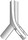
Endobronchial
intubation
occurs if too long a tube is used and inserted into one
of the mainstem bronchi. The un-intubated lung does not
contribute to gas exchange and the large volume of blood
flowing through this lung results in a substantial right
to left shunt.
Signs
are those of arterial hypoxemia, including cyanosis and
labored breathing. In addition, uptake of the inhalation
anesthetic agent may be impaired, resulting in an unexpectedly
light plane of anesthesia.
This
problem may be avoided by trimming of the tube to the
correct length and securing it firmly around the muzzle
or lower jaw of the patient. If too long a tube is used
and it is tied around the back of the patient's neck,
movement of the tube during surgery may move the tip of
the tube into a bronchus.
|
 |
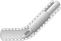
| Impaction
of the tip of the tube against the tracheal wall may result
in respiratory obstruction, particularly where the trachea
contains a sharp bend, such as the thoracic inlet. |
 |
|
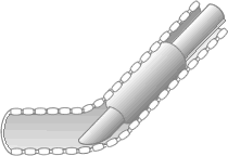
The Murphy eye, incorporated into many modern tubes,
permits airflow to take place even if this has occurred. |
 |
| Herniation
of the cuff over the lumen of the tube may happen if
the cuff of an old, perished tube is over-inflated. This,
again, will cause respiratory obstruction. |
 |
| Compression
of the lumen of the tube by the cuff may be caused by
over-inflation of the cuff or by gradual diffusion of nitrous
oxide onto the cuff during the course of anesthesia.This
problem is more common when silicone rubber tubes are used. |
 |
| Stretching
of the tracheal wall may be caused by over-inflation
of the cuff. This may lead to tracheitis, pressure necrosis
of the tracheal wall, or tracheal rupture. |
 |
|
Fires are a danger associated with the increasing
use of lasers for airway and oral surgery. Steps which
may be taken to reduce this extremely serious hazard
include:
-
Using
special laser tubes, which may be made of jointed
metal or clear plastic (with no radiopaque strip).
- Wrapping
exposed portions of the tube with aluminum tape.
- Use
of helium-oxygen mixtures which are less supportive
of combustion than oxygen alone or oxygen-nitrous
oxide mixtures.
|
Laryngeal
masks 
|
